Posterior & Anterior Crossbite: Diagnosis and Correction with Clear Aligners
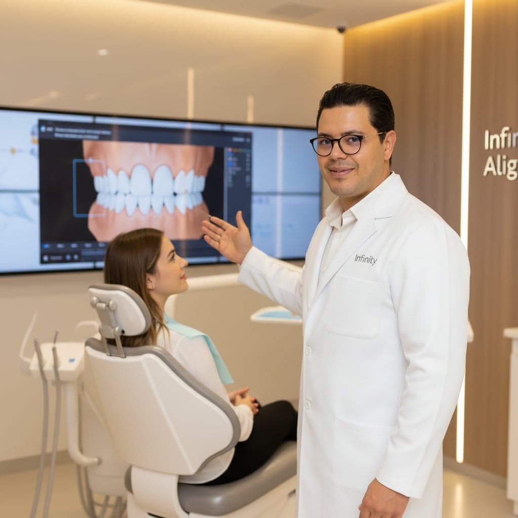
Technical clinical article on crossbite differential diagnosis and treatment with clear aligners: classification, biomechanics, attachments, staging, and retention protocols.
Crossbite is defined as an aberrant buccolingual relationship between one or more maxillary and mandibular teeth in centric occlusion. Specifically, a maxillary tooth occludes lingual to the corresponding mandibular antagonist — the inverse of the normal relationship described by Angle and refined in the Andrews Six Keys. Beneath this apparent positional simplicity often lie underlying skeletal, functional or eruptive disturbances. For the clinician evaluating an aligner indication, accurate aetiologic classification of the crossbite is the single most decisive diagnostic step.
Untreated — particularly when functional in origin — crossbite can lead to asymmetric condylar remodelling, accelerated periodontal attachment loss on involved teeth, and persistent mandibular asymmetry in growing patients. This article details classification, differential diagnosis, aligner candidacy, adapted biomechanics, and the retention protocol.
1. Classification and Epidemiology
Crossbites are classified along three axes: location (anterior vs posterior), laterality (uni- vs bilateral), and origin (dental, skeletal, or functional). Reported prevalence in the permanent dentition ranges from 7% to 23% depending on the population studied, with posterior crossbite considerably more common than anterior crossbite in non-syndromic populations.
1.1 Anterior Crossbite
One or more maxillary incisors occlude lingual to the mandibular incisors. Prevalence: 4–5% in the mixed dentition. Must be carefully differentiated from true skeletal Class III malocclusion.
1.2 Posterior Crossbite
Maxillary posterior teeth occlude lingual to mandibular antagonists. Prevalence: 8–22%. Most commonly unilateral with a functional shift; also the most under-diagnosed form in adult populations.
2. Aetiology — Skeletal vs Dental vs Functional
The aetiologic factor dictates the treatment ceiling. A purely dental crossbite — tipped teeth on an otherwise symmetric base — is the most aligner-amenable presentation. A skeletal crossbite requires expansion of the apical base and, in most adult cases, lies beyond the biomechanical envelope of aligners alone.
2.1 Dental aetiology
- Localized arch length deficiency with palatal eruption of laterals or canines.
- Persistent deciduous teeth altering eruption path of the successor.
- Supernumerary or impacted teeth deflecting normal eruption.
- Iatrogenic — over-contoured restorations, residual scalloped contacts.
2.2 Skeletal aetiology
- Maxillary transverse hypoplasia — most common skeletal cause.
- Mandibular transverse excess — comparatively rare in non-syndromic patients.
- Pierre Robin sequence, cleft lip/palate, and other developmental syndromes.
- Habitual mouth breathing and low tongue posture leading to a narrow palatal vault during development.
2.3 Functional aetiology
The functional shift is diagnostically pivotal. The patient's mandible may be symmetric in centric relation but deflect 1–3 mm laterally to engage in maximum intercuspation. Failure to recognize this shift leads to over-treatment of a perceived skeletal asymmetry that is, in fact, dento-alveolar. Clinical rule: always evaluate the patient with the mandible guided into centric relation before finalizing the transverse diagnosis.
3. Posterior Crossbite
Posterior crossbite involves the premolars and molars and is the more biomechanically demanding presentation when planning aligner therapy. The most common form — unilateral with a functional shift — is also the most under-diagnosed in adult populations who have learned to ignore the bite asymmetry.
3.1 Unilateral with functional shift
Clinical signature: symmetric arches and condyles in centric relation, deviation of the mandibular midline on closure, audible occlusal interference on the involved side. The crossbite resolves bilaterally when occlusion is examined in centric. Aligner treatment requires elimination of the interference (selective IPR, posterior intrusion, or buccal expansion) so the mandible auto-corrects to centric.
3.2 Bilateral posterior crossbite
Bilateral involvement strongly suggests skeletal maxillary deficiency. In adults, dento-alveolar expansion of more than 3–4 mm via aligners alone introduces unfavourable buccal crown tipping with limited apical base movement. Surgically-assisted rapid palatal expansion (SARPE) or mini-screw-anchored expansion (MARPE) is usually required prior to or in parallel with aligner therapy.
Biological limit: in skeletally mature patients, sustainable buccal dento-alveolar expansion via clear aligners is generally limited to 2–3 mm per quadrant. Beyond this threshold, the risk of buccal bone dehiscence, gingival recession, and relapse rises sharply. Plan expansion goals in millimeters per side, never as a global arch-width target.
4. Anterior Crossbite
Anterior crossbite involves the incisors and is often misdiagnosed as early Class III malocclusion. The distinction is critical: pseudo Class III (functional anterior crossbite) is well within the aligner envelope, while true skeletal Class III is not.
4.1 Single-tooth anterior crossbite
A single maxillary incisor in linguoversion, with adequate arch length distal to the crossbite and a normal overjet on the remaining incisors. Excellent aligner candidacy: 12–20 stages, with attachments controlling labial root torque, and IPR distal to the involved tooth if required.
4.2 Multi-tooth anterior crossbite
Two or more incisors in crossbite raises the index of suspicion for skeletal involvement. Cephalometric ANB, Wits appraisal, and overjet measurement guide the differential between dental and skeletal causes. A negative ANB beyond −1.5° or a Wits beyond −2 mm typically exceeds the aligner-only envelope in non-growing patients.
5. Clinical Diagnosis
A reproducible diagnostic protocol prevents the most common downstream treatment failures. The Infinity Aligner clinical team recommends the following sequence:
- Extraoral evaluation: chin point relative to facial midline at rest, on smile, and on functional closure. Lower facial third symmetry. Lip competence.
- Functional analysis: guide the patient into centric relation. Identify the first point of contact, evaluate the slide and any laterodeviation into maximum intercuspation.
- Intraoral exam: map the crossbite teeth. Assess buccal bone, gingival biotype, and existing recession or fenestrations on involved roots.
- Periodontal assessment: probing depths, attachment levels, and recession measurements at every crossbite tooth. Periodontal stability is non-negotiable before expansion.
- Digital records: intra-oral scan, panoramic radiograph at minimum, lateral and PA cephalogram when skeletal involvement is suspected. CBCT for borderline expansion cases.
- Treatment objectives: define corrected tooth positions, expansion goals in millimeters per quadrant, and inter-arch coordination before submitting to the planning portal.
6. Imaging and Records
Two-dimensional imaging remains adequate for most posterior dental crossbites. Three-dimensional CBCT is indicated when buccal expansion of more than 2 mm per side is planned, when the apical base is suspected to be narrow, when fenestrations are detected clinically, or when impacted teeth contribute to the malocclusion.
| Record | When indicated | Clinical contribution |
|---|---|---|
| Intra-oral scan | All cases | Arch form, midlines, tooth size, occlusal contacts |
| Panoramic radiograph | All cases | Root parallelism, impacted teeth, periapical pathology |
| Lateral cephalogram | Anterior crossbite + suspected skeletal Class III | ANB, Wits, incisor inclination, sagittal jaw relation |
| PA cephalogram | Bilateral posterior crossbite, asymmetry | Maxillo-mandibular transverse discrepancy |
| CBCT | Expansion > 2 mm/side, fenestrations, impactions | Buccal cortical bone thickness, root proximity |
| Photographs | All cases | Facial symmetry, smile arc, incisal exposure |
7. Differential Diagnosis
Three differentials govern aligner candidacy: pseudo vs true Class III, dental vs skeletal transverse deficiency, and functional shift vs true mandibular asymmetry. Each is resolved by combining the centric-relation closure test, cephalometric measurement, and CBCT inter-molar width assessment.
- Pseudo Class III: anterior crossbite, neutral ANB, normal incisor inclination, anterior interference forces protrusion → aligner candidate.
- True Class III: negative ANB, deficient maxilla and/or excessive mandible, no functional shift → orthognathic surgical referral.
- Functional shift: symmetric CR, asymmetric maximum intercuspation, deviation on closure → aligner candidate (eliminate the interference).
8. Aligner Candidacy Matrix
The matrix below summarizes our internal candidacy framework. It is not a substitute for clinician judgment but provides a defensible baseline when triaging consults:
| Candidacy | Typical indications |
|---|---|
| Excellent | Single-tooth anterior crossbite · unilateral posterior dental crossbite < 2 mm · pseudo Class III without skeletal divergence |
| Good | Multi-tooth anterior crossbite, neutral ANB · unilateral posterior crossbite 2–3 mm · functional shift requiring interference elimination |
| Borderline | Bilateral posterior crossbite ≤ 3 mm/side in young adult, no recession · mild skeletal divergence on PA ceph |
| Refer | True skeletal Class III · bilateral crossbite > 4 mm/side · existing recession or buccal dehiscence · syndromic patients |
9. Aligner Biomechanics
Clear aligners apply force through tooth-contacting plastic deformation. Crossbite correction relies on three primary movements — buccal crown translation, root torque, and intrusion of interfering cusps. Each requires deliberate attachment design and staging to be predictable.
9.1 Force vectors and attachment design
- Optimized rectangular attachment on the buccal surface of premolars and molars for buccal expansion; orientation tracking crown rather than apex movement.
- Horizontal beveled attachment on the labial surface of crossbite incisors to deliver labial root torque and avoid pure crown tipping.
- Bite ramps on the palatal surface of maxillary central incisors to open the bite and disclude posterior interferences during posterior expansion.
- Power ridges as a torque adjunct when attachment placement is contraindicated (visible anterior aesthetics).
9.2 Movement velocity
Predictable per-stage movement targets validated at the lab:
- Buccal crown translation — 0.20 mm per stage with optimized attachment.
- Labial root torque — 1.5° per stage with horizontal beveled attachment.
- Posterior intrusion (interference) — 0.15 mm per stage with bilateral support.
- Anterior labial tipping (single tooth) — 1.0 to 1.5° per stage.
10. Staging and Force Control
The Infinity Aligner planning portal supports staging at 1- or 2-week intervals. For crossbite cases, the following sequencing rules apply:
- Stages 1–3 (alignment): eliminate rotations and minor crowding. Stabilize the arches before transverse work begins.
- Stages 4–8 (interference removal): intrude or distalize teeth that prevent the mandible from reaching centric. The functional shift, if present, often resolves spontaneously by stage 7.
- Stages 8–18 (transverse correction): buccal expansion with bilateral attachments to prevent posterior open bite. Symmetric movement is essential.
- Stages 18–24 (overcorrection): 1.5× the desired final position to compensate for relapse during retention.
- Stages 24–end (detailing): final tip, torque, and interdigitation refinements before retention.
Bilateral mechanics rule: for any unilateral transverse correction exceeding 1 mm, place a counter-balancing attachment on the contralateral side to prevent unwanted dental midline drift. Even when only one side is in crossbite, the mechanics should always be considered bilateral.
11. Illustrative Clinical Case
A 34-year-old female patient presented with unilateral right posterior crossbite from canine to second molar, 2.5 mm transverse deficiency on the right side, and a 1.8 mm functional shift to the right on closure. Centric relation closure was symmetric. ANB 2.4°, no skeletal asymmetry on PA cephalogram.
Plan approved on day 14: 28-stage aligner plan. Optimized attachments on UR3, UR4, UR5, UR6, UR7. Counter-balancing on UL4 and UL6. Bite ramps on UR1 and UL1. Compliance: 22 hrs/day. Functional shift resolved at stage 11. Crossbite corrected at stage 19. Stages 20–28: detailing and overcorrection. Outcome at month 9: symmetric Class I posterior occlusion, coincident midlines in centric, 1.5 mm overjet, 2.0 mm overbite. No recession or root resorption on post-treatment CBCT. Stability confirmed at the 12-month recall.
12. Retention Protocol
Transverse corrections relapse more aggressively than alignment corrections. The retention protocol following crossbite treatment must include:
- Vacuum-formed retainer full-time × 6 months post-treatment, then nightly indefinitely.
- Bonded lingual retainer where rotational relapse risk is high (canines, lower incisors).
- Annual occlusal screening for functional disturbances during the first 3 years.
- Patient education: retention is lifelong. Communicate this at the consultation, not at the end of treatment.
13. Conclusion
Clear aligner therapy has become a clinically defensible primary modality for the majority of dental crossbites — single-tooth anterior crossbites, unilateral posterior crossbites with functional shift, and selected mild bilateral crossbites in periodontally sound patients. The key clinical decisions are made before the first aligner is delivered: an accurate functional and skeletal differential, deliberate attachment design, bilateral mechanics, and a retention plan that respects the elastic memory of the periodontal ligament.
Cases that exceed the aligner envelope — true skeletal Class III, transverse deficiencies greater than 3 mm per quadrant, or compromised periodontal support — remain firmly in the territory of orthognathic surgery, surgically-assisted expansion, or specialist orthodontic referral. Recognizing those cases at the consultation is the most valuable diagnostic skill a general dentist can develop.
Infinity Aligner
Clinical & editorial team
More articles
How to Choose Clear Aligners in Tunisia
Clear Aligners vs Braces: Complete Comparison
How Much Do Clear Aligners Cost in Tunisia?
How Long Does Clear Aligner Treatment Take?
How to Clean and Care for Your Clear Aligners Daily

Clear Aligners for Teenagers: What Parents Need to Know
Gap Teeth & Diastema: Can Clear Aligners Fix Them?
Can You Play Sports with Clear Aligners?

3D Smile Simulation: How It Works and What to Expect

Orthodontic Relapse: Why Teeth Move After Treatment
Intraoral Scanner vs Traditional Impressions: What's the Difference?
Clear Aligners for Adults: Is It Ever Too Late?
Wisdom Teeth and Clear Aligners: Are They Compatible?

Clear Aligners: A Versatile Therapeutic Approach in Orthodontics

Orthodontics and Periodontal Health: What Every Patient Needs to Know
The Technology Behind Clear Aligners: From Design to Tray

How to Prepare for Your Orthodontic Consultation: 10 Questions to Ask
Bruxism and Clear Aligners: Can You Correct and Protect at the Same Time?

Class II Malocclusion and Clear Aligners: What Treatment Options Exist?
Clear Aligners and Dental Implants: Protocol, Timing and Clinical Cases
Digital Workflow and Aligner Biomechanics: The Advanced Practitioner Protocol
The Limits of Clear Aligners: Identifying Complex Cases Beyond Their Reach
Aligner Manufacturing: Thermoforming, 3D Printing and Polymer Science
Invisible Orthodontics in 2030: Predictive AI, Direct Printing and Active Materials
Smile Without Borders: The Price of Aligners Between Tunisia, Europe and Canada
Attachments in Invisible Orthodontics: The Silent Levers of Biomechanics

The Smile Beyond the Mirror: The Profound Impact of Dental Alignment on Psychological Well-being
Tunisia: The New Eldorado of Digital Dentistry and Medical Tourism
Post-Orthodontic Retention: The Forgotten Step That Decides Everything
What You Can (and Cannot) Eat With Clear Aligners
Teeth Whitening and Clear Aligners: Can You Treat and Whiten at the Same Time?
TMJ and Clear Aligners: How Invisible Orthodontics Acts on Jaw Pain
The Different Types of Dental Retainer: A Complete Guide After Orthodontic Treatment
The hidden dangers of metal braces: what patients need to know
Root resorption: the silent risk of poorly managed orthodontic treatments
Orthodontics and cavities: how metal braces compromise dental hygiene
Orthodontic relapse: why teeth return to their original position
Braces and chronic pain: the underestimated impact on daily quality of life
Intraoral scanner vs conventional impressions: dimensional analysis and clinical implications
CAD/CAM digital workflow in orthodontics: from intraoral scanning to aligner fabrication
Deformation, shrinkage and cumulative errors: why the conventional impression chain compromises aligner precision
Intraoral Scanners 2025: Complete Technical and Metrological Comparison
Why Choose Infinity Aligner? The Complete Guide for Dental Practitioners
Composite Selection for Aligner Attachments: Mechanical Analysis and Clinical Protocol
TMJ and Malocclusion: Differential Diagnosis and Clinical Approach for the Practitioner
Travelling with Clear Aligners: The Complete Guide for Stress-Free Trips
The First Month with Aligners: What to Expect Week by Week
Pre-Prosthetic Orthodontics: Preparing the Site Before Implants or Veneers
How to Present Aligners to a Hesitant Patient — A Consultation Script
Anterior Open Bite and Aligners: Protocol, Intrusion and Relapse Risk
Tobacco, Cigarettes and Clear Aligners: Everything You Need to Know
Invisalign vs Infinity Aligner: Complete Price Comparison in Tunisia (2026)
What Happens If You Don't Wear Your Aligners 22 Hours a Day?
Myths and Misconceptions About Clear Aligners: What Patients and Dentists Get Wrong
Sleep Apnea and Orthodontics: The Dentist's Role in Screening and Management
Consult a certified dentist
Our 200+ certified partners in Tunis, Sfax, Sousse and the francophone zone welcome you.
Book a consultation