Static Surgical Guides: Tooth-, Mucosa- or Bone-Supported — Which Guide for Which Case?
The tissue a surgical guide rests on determines its stability, its access to the surgical site and, according to systematic reviews, part of its accuracy. Three support families, what three literature reviews report, and a selection grid by type of edentulism.
A static surgical guide transfers the implant plan made on the CBCT and the digitised dental surface to the chairside: virtually positioned guide sleeves dictate the axis and depth of drilling. This transfer is only reliable if the guide returns to exactly the same place every time. The structure it rests on — its support — therefore determines its stability, the surgical approach and part of the final error. This article compares the three support families, summarises what three systematic reviews published in Clinical Oral Implants Research allow us to say, and proposes a selection grid. It is written for practitioners: it replaces neither training in the guided surgery system used nor clinical judgement.
1. Three Support Families
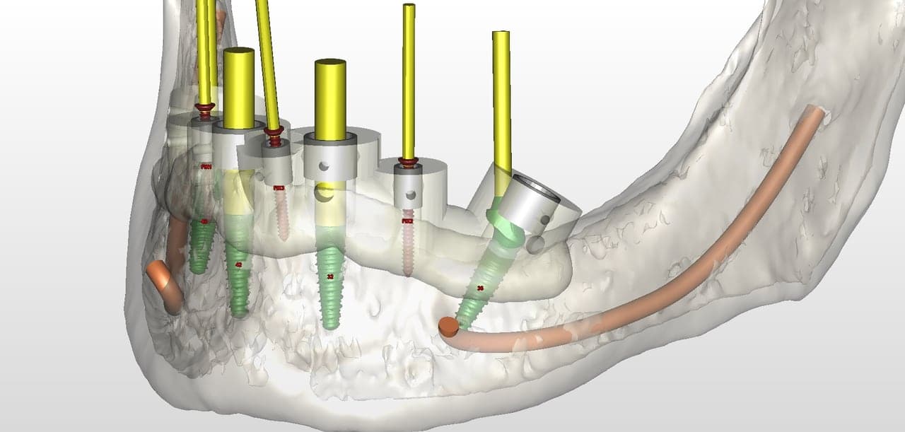
Guides are classically divided into tooth-, mucosa- and bone-supported, to which are added mixed guides (for example teeth and mucosa in an extended partial edentulism). On the header illustration, a mandibular plan, the translucent guide body carries sleeves (white cylinders) aligned with the axis of each implant (green, extended in yellow), the course of the inferior alveolar nerve is traced in orange and three fixation pins (markers FIX1 to FIX3) anchor the guide in the bone. This is the logic shared by all three families: a guide only guides well if it is immobilised.
| Support | Typical situation | Strengths | Points of caution |
|---|---|---|---|
| Tooth | Partial edentulism with numerous, stable remaining teeth | Unambiguous seating on rigid landmarks; flapless approach possible; inspection windows to check seating | Distribution of teeth along the arch; access and length of instruments in the posterior region; mobile teeth or bulky restorations to check |
| Mucosa | Complete or very extended edentulism, adequate ridge, flapless surgery | Minimally invasive: no flap, simpler postoperative course | Compressible, mobile mucosa; indirect scan data (scan prosthesis, markers); fixation pins strongly recommended; no view of the bone |
| Bone | Complete or extended edentulism with irregular ridge, regularisation osteotomy | Rigid seating on bone; direct view of the bone; ridge regularisation in the same session | Flap mandatory (more invasive); planning on the final bone level; guide placed on exposed bone, so rigorous fixation |
2. What the Systematic Reviews Report
Three reviews published in the same journal between 2009 and 2018 structure the available data on static guides. Their conclusions complement rather than contradict each other.
| Review | Data | What it reports |
|---|---|---|
| Schneider et al., 2009 (EAO consensus conference) | Eight accuracy studies | Mean deviation of 1.07 mm at the entry point and 1.63 mm at the apex; maximum deviations clearly higher than the means; no significant difference by production method or type of support at that date |
| Raico Gallardo et al., 2017 | Eight clinical studies, four in the meta-analysis | Bone-supported guides deviate more than tooth-supported guides (angle, entry point, apex), but the entry-point and apex difference disappears when only retrospective studies are analysed; mucosa-supported guides deviate less than bone-supported guides; no difference between mucosa- and tooth-supported guides |
| Tahmaseb et al., 2018 (ITI consensus conference) | 20 studies, 2,238 implants, 471 patients | Mean deviation of 1.2 mm at the entry point (95% CI: 1.04–1.44), 1.4 mm at the apex (1.28–1.58) and 3.5° in angle (3.0–3.96); better accuracy in partial than in complete edentulism; a safety margin of at least 2 mm should be respected |
Three practical lessons. First, the order of magnitude: a static guide is not accurate to a tenth of a millimetre, and the 2 mm safety margin called for by Tahmaseb et al. is not a theoretical precaution. Second, support matters: the 2017 review concludes that the supporting tissue influences accuracy, with tooth and mucosa support outperforming bone support in the pooled analysis — an advantage that the analysis restricted to retrospective studies does not find for the entry point and apex. Third, the type of support is linked to the type of edentulism: tooth support is mostly met in partial edentulism, mucosa and bone support mostly in complete edentulism, where accuracy is lower. The two effects cannot therefore be separated in the data; this reading is our own inference, not a finding of these reviews.
3. Which Support for Which Case: A Decision Grid
The grid below summarises the usual logic. It replaces neither case-specific planning nor the recommendations of the manufacturer of the system used.
| Clinical situation | Preferred support | Point of attention |
|---|---|---|
| Short partial edentulism (1 to 3 teeth), stable neighbouring teeth | Tooth | Scan the whole arch; check seating on several teeth before anaesthesia |
| Extended partial edentulism, few remaining teeth or free posterior distal area | Tooth or mixed (teeth + mucosa) | Short, poorly distributed tooth support: add fixation and widen the safety margin |
| Complete edentulism, regular ridge, sufficient keratinised gingiva, flapless surgery | Mucosa with fixation pins | Scan prosthesis and radiopaque markers; seating check; lower accuracy than in partial edentulism in the 2018 review |
| Complete edentulism, irregular ridge or planned regularisation osteotomy | Bone (with flap) | Plan on the bone level after regularisation; rigid fixation; plan the surgical exposure |
4. Securing the Seating: Seven Checks Before Operating
- Try the guide on the printed model or in the mouth before anaesthesia, using the inspection windows where they exist
- Fix the guide with pins or screws whenever the support is mucosa or bone, and check after each drill pass that it has not moved
- Check the fit after sterilisation or storage: a deformed guide must be remade, not forced
- Check data consistency: in complete edentulism, the scan prosthesis must be perfectly stable in the scanner and the radiopaque markers correctly recognised by the software
- Measure mouth opening and available height: sleeve, key and drill must fit in the space, especially in the posterior region
- Respect a safety margin of at least 2 mm around the inferior alveolar nerve, the sinus and neighbouring teeth (Tahmaseb et al., 2018)
- Keep a fallback plan: know how to continue freehand or postpone if the guide does not seat, then check the actual position on the postoperative CBCT to document the deviation
5. Limits of the Data
The three reviews stress the heterogeneity of techniques, software, manufacturing processes and study designs. The 2018 review rests on a single randomised controlled trial, eight uncontrolled retrospective studies and eleven uncontrolled prospective studies. These works mainly measure the accuracy of implant position, not long-term survival or patient satisfaction, and their means cannot be transposed as such to a given system, printer or operator. Systematically superimposing the postoperative CBCT on the plan remains the best way to know one’s own accuracy.
Key Takeaways
- The support of the guide determines its stability, the surgical approach and part of the accuracy.
- Tooth support: preferred when the remaining teeth are sufficiently numerous, stable and well distributed.
- Mucosa support: minimally invasive, but subject to mucosal mobility; fixation and seating checks are essential.
- Bone support: rigid seating and a view of the bone, at the cost of a flap.
- Whatever the family, plan a safety margin of at least 2 mm and check the position obtained.
References
- Schneider D, Marquardt P, Zwahlen M, Jung RE. A systematic review on the accuracy and the clinical outcome of computer-guided template-based implant dentistry. Clin Oral Implants Res. 2009;20(Suppl 4):73–86.
- Raico Gallardo YN, da Silva-Olivio IRT, Mukai E, Morimoto S, Sesma N, Cordaro L. Accuracy comparison of guided surgery for dental implants according to the tissue of support: a systematic review and meta-analysis. Clin Oral Implants Res. 2017;28(5):602–612. doi:10.1111/clr.12841
- Tahmaseb A, Wu V, Wismeijer D, Coucke W, Evans C. The accuracy of static computer-aided implant surgery: a systematic review and meta-analysis. Clin Oral Implants Res. 2018;29(Suppl 16):416–435. doi:10.1111/clr.13346
Editorial note
This article is written for scientific and professional monitoring purposes. The studies cited are drawn from peer-reviewed publications. Infinity Aligner does not endorse the results of third-party studies and recommends that professionals consult the original publications for any clinical application.
Infinity Aligner — Scientific team
Technology watch & dental literature review
More analyses
Mar 2026
Photobiomodulation (LLLT) and Bone Healing in Implantology: Clinical Evidence, Dosimetry Protocols and 2024 Post-surgical Applications
Mar 2026
Osteodensification and Bone Condensation in Implantology: Versah Technique, Condensation Osteotomes and Bone Quality Improvement Protocols in D3–D4 Bone
Mar 2026
Peri-Implantitis: Diagnosis, Implant Surface Decontamination and Surgical Bone Regeneration — Evidence-Based Protocols 2024
Integrate innovation into your practice
Join the Infinity Aligner network and access the most advanced digital tools for your patients.
Become a partner