Complete Digital Workflow for All-on-X: From Virtual Planning to Same-Day Prosthetics
The complete digital workflow — CBCT + optical impression + virtual planning + printed surgical guide + pre-milled provisional prosthesis — now makes it possible to complete a full All-on-X rehabilitation in a single surgical day with millimetre precision. Protocol, software and clinical validation.
"Teeth in a Day" has long been the marketing promise of the All-on-4 protocol. In 2024, thanks to the convergence of the complete digital workflow — pre-operative CBCT, intraoral scanner, 3D surgical planning software, printed surgical guide, pre-milled provisional prosthesis — this promise has become a daily clinical reality in implant centres of excellence. Moreover, the digital workflow enables delivery of a dimensionally precise provisional prosthesis the day after surgery, and a definitive prosthesis within 6 to 8 months, without a physical impression at any stage of the protocol.
1. Steps of the Complete All-on-X Digital Workflow
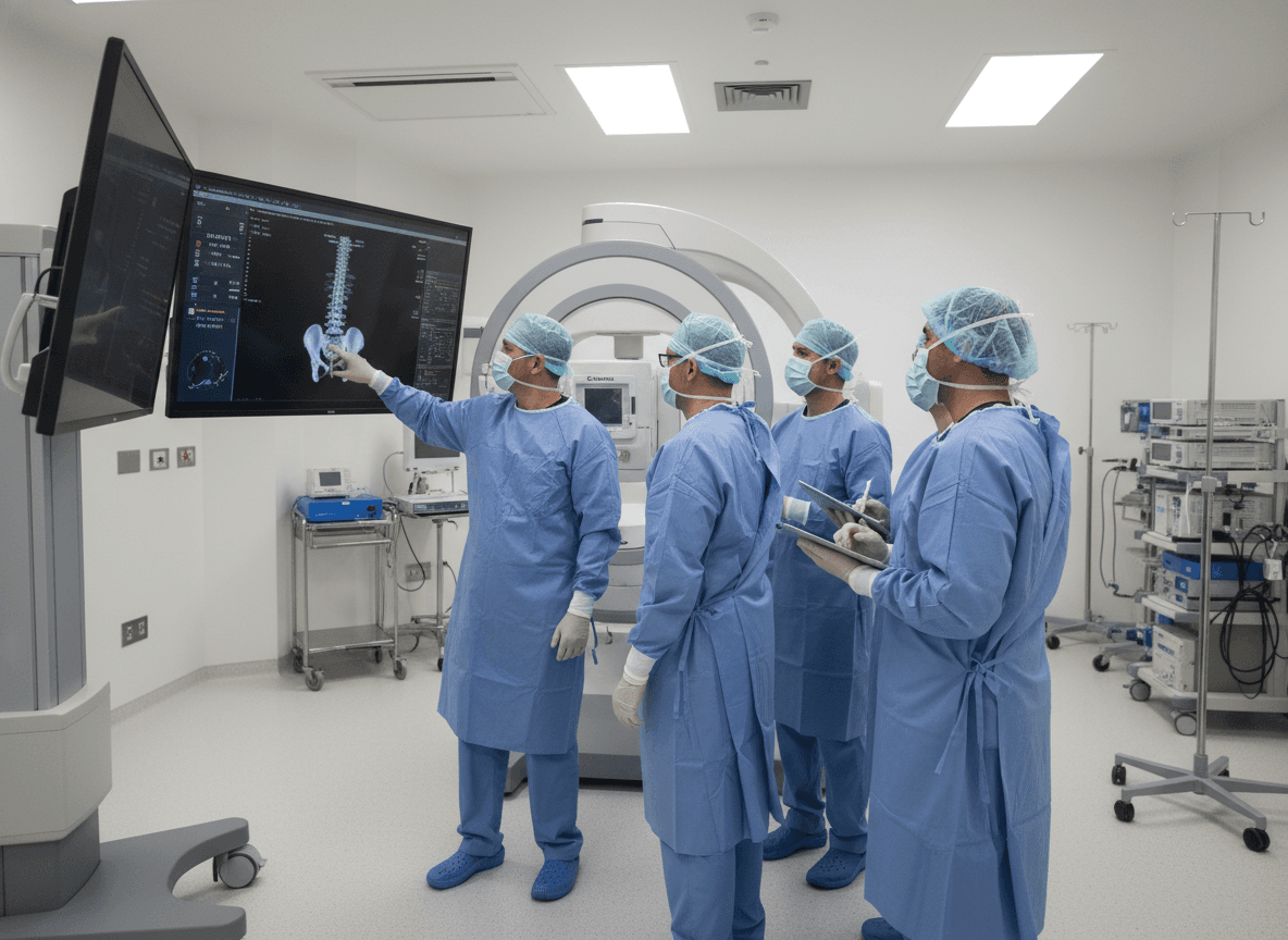
- Step 1 — CBCT assessment (D-30 to D-7): large field volumetric acquisition (16 × 13 cm), DICOM format export for import into planning software
- Step 2 — Intraoral scan (D-30 to D-7): acquisition of current dental/prosthetic situation, gingival profile and current VDO. CBCT + STL scan fusion in software
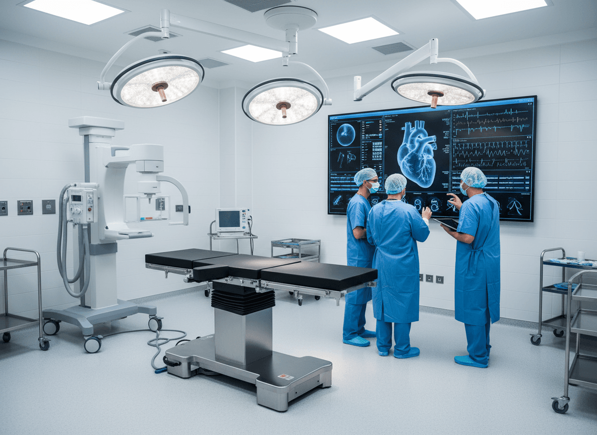
- Step 3 — Digital Smile Design (DSD): design of final aesthetic project, incisal edge position, occlusal parameters. Prosthetic design guides implant planning
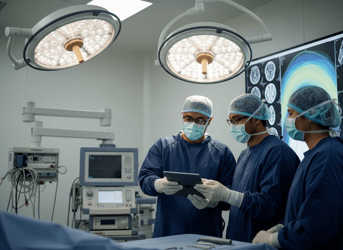
- Step 4 — Virtual planning (dedicated software): 3D implant positioning, AP spread calculation, anatomical safety margin verification, cantilever biomechanical simulation
- Step 5 — Surgical guide printing (D-7 to D-3): stereolithographed guide (Class IIa CE resin), virtually validated, autoclave B sterilisable
- Step 6 — Provisional prosthesis pre-milling (D-7 to D-3): reinforced PMMA milling based on DSD data and planned MUA abutment positions — without physical impression
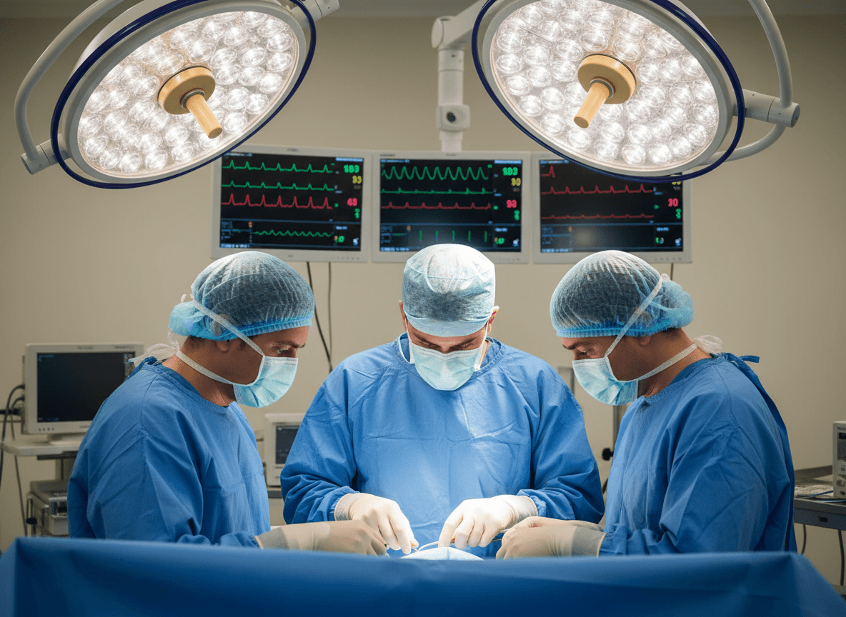
- Step 7 — Guided surgery (D0): flapped or flapless procedure depending on bone availability, implant placement via guide, MUA abutment connection, intraoperative intraoral scan on MUA transfer keys
- Step 8 — Adjustment and delivery (D0 or D1): screwing of pre-milled prosthesis onto abutments, final occlusal adjustment, patient instructions
2. Reference Software: 2024 Comparison
| Software | Publisher | Strengths | Guide integration | Integrated prosthetic workflow |
|---|---|---|---|---|
| coDiagnostiX | Dental Wings / Straumann | Academic reference, exhaustive implant library | Yes (SLA/DLP) | Partial (STL export) |
| Simplant Pro | Dentsply Sirona | Integrated FEA, load simulation | Yes | Partial |
| Implant Studio | 3Shape | Intuitive interface, native TRIOS workflow | Yes (SLA/DLP) | Yes (3Shape Dental System link) |
| Blue Sky Plan | Blue Sky Bio (free) | Free, open source compatible | Yes (STL export) | No |
| RealGUIDE | 3Diemme | Integrated dynamic navigation | Yes + navigation | Partial |
| Nobel Clinician | Nobel Biocare | Native All-on-4 integration, turnkey service | Yes (bureau service) | Yes (NIRI bridge) |
3. Guided Flapless Surgery: Advantages and Limitations
Flapless surgery — without a mucoperiosteal flap — is possible in the All-on-X protocol when bone volume is sufficiently predictable (high-resolution CBCT) and the surgical guide is mucosal or implant-supported. Its advantages are documented: operating time reduction of 20–35%, significantly reduced intraoperative bleeding, lower post-operative pain (VAS at D1: 3.2 vs 5.1 for flap surgery — JOMI 2023 study), faster return to eating. Its limitations: requires high-resolution CBCT (voxel ≤ 100 µm), is not indicated if levelling osteoplasty is needed, does not allow direct bone visualisation in the event of an unforeseen intraoperative complication. The flapless vs open flap decision must be made case by case after rigorous CBCT analysis — not as a systematic policy.
4. Digital Workflow Accuracy: 2024 Validation Data
The accuracy of the complete digital workflow (planning → guided surgery → pre-milled prosthesis) is the limiting factor in result quality. A multicentre prospective study published in the International Journal of Implant Dentistry (2024) on 312 All-on-X implants placed under static tooth-mucosal guide and post-operatively CBCT-scanned documents the following mean deviations: angular error 1.9 ± 1.1°, entry error 0.51 ± 0.28 mm, apical error 0.61 ± 0.34 mm. These values are clinically acceptable for the vast majority of cases. For high-risk anatomical zones (mandibular nerve proximity < 2 mm, thin sinus wall), dynamic navigation (Image-Guided Surgery — RealGUIDE or X-Guide system) offers superior precision: apical error 0.36 ± 0.18 mm documented in the same study.
5. Integration into Tunisian Practice: Current State and Prospects
In Tunisia, adoption of the complete digital workflow for All-on-X rehabilitations is rapidly progressing in advanced private implantology clinics. The main identified barriers are equipment investment (CBCT, intraoral scanner, 3D printer, planning software: total investment of DT 150,000–300,000 for a complete setup) and the learning curve for care teams. The first Tunisian centres to have integrated this protocol are documenting significantly higher patient satisfaction, reduced intraoperative complications and result standardisation that reinforces the confidence of international patients in the high-quality dental medical tourism model.
Editorial note
This article is written for scientific and professional monitoring purposes. The studies cited are drawn from peer-reviewed publications. Infinity Aligner does not endorse the results of third-party studies and recommends that professionals consult the original publications for any clinical application.
Infinity Aligner — Scientific team
Technology watch & dental literature review
More analyses
Mar 2026
Photobiomodulation (LLLT) and Bone Healing in Implantology: Clinical Evidence, Dosimetry Protocols and 2024 Post-surgical Applications
Mar 2026
Osteodensification and Bone Condensation in Implantology: Versah Technique, Condensation Osteotomes and Bone Quality Improvement Protocols in D3–D4 Bone
Mar 2026
Peri-Implantitis: Diagnosis, Implant Surface Decontamination and Surgical Bone Regeneration — Evidence-Based Protocols 2024
Integrate innovation into your practice
Join the Infinity Aligner network and access the most advanced digital tools for your patients.
Become a partner